Training Principles
Strength training, also known as resistance or weight training has the greatest potential benefit on both your performance as an athlete and overall heath.
One can think of exercise as medicine, since it has the potential to lower the risk of nearly all chronic diseases.
The problem is key principles of training and acute program variables are often misunderstood, leaving people unaware of what the effective ‘dose’ is. (1,2)
Another issue is that untrained individuals are highly adaptable, which means almost any exercise program, load and method may increase strength in the short term. This is likely due to neural adaptations in response to the new training stimulus and will have a low ceiling for additional improvements. If you wish to pursue further strength adaptations, progressive overload is essential. (2)

The capability of a strength training program to achieve a specific or desired outcome depends on two things: (3-5)
Following the key principles of training
Manipulating acute program variables
Key Principles of Training:
Overload = exposure of tissues to greater than accustomed to training stress
Reversibility = removal of tissue loading results in loss of adaptations
Progression = continuous and organized increases in training stress or overload to enhance or maintain training adaptations
Individualization = modification of training due to different variations in the response to training
Periodization = planned organization and modification of training programs over time to optimize progression
Specificity = matching training to the movements patterns and intensities that occur during sport to enhance performance
Acute Program Variables:
Intensity
Volume
Rest
Frequency
Muscle action
Exercise selection & order
Intensity:
Intensity or load refers to the amount of weight assigned to an exercise set and is the most important variable in programming to drive adaptation. Intensity can be determined by:
-Weight
-% of RM (Repetition Maximum)
-Rating of Perceived Exertion
-Repetitions in Reserve
Volume:
Volume can describe the number of repetitions per set, number of sets per session, or the number of sessions per week
Rest:
Rest is the time allotted for recovery between sets and exercises. Rest is the second most important program variable, since it will determine the intensity and demand of the exercise. The length of the rest period is dependent on the training goal, the relative load lifted, and the training status of the individual.
Frequency:
Training frequency refers to the number of training sessions completed in a given time period, usually over a week. Frequency is dependent on the type of training, the status and recovery ability of the individual.
Muscle action:
An exercise can occur with the muscle shortening (concentric), lengthening (eccentric) or not moving (isometric). Most exercises involve all three muscle actions. Though some specific exercises may focus on one of the three muscle actions.
Exercise selection & order:
There are an endless number of exercises that can be utilized while training. Typically, multi-joint exercises (i.e. Squat, deadlift, bench press) are performed first, as they are more intense and require higher muscle activation and energy expenditure. Followed by single-joint exercises as required.
-
1. Hansford, H. J., Wewege, M. A., Cashin, A. G., Hagstrom, A. D., Clifford, B. K., McAuley, J. H., & Jones, M. D. (2022). If exercise is medicine, why don't we know the dose? An overview of systematic reviews assessing reporting quality of exercise interventions in health and disease. British journal of sports medicine, 56(12), 692–700.
2. Maestroni, L., Read, P., Bishop, C., Papadopoulos, K., Suchomel, T. J., Comfort, P., & Turner, A. (2020). The Benefits of Strength Training on Musculoskeletal System Health: Practical Applications for Interdisciplinary Care. Sports medicine, (50(8), 1431–1450.
3. Bird, S. P., Tarpenning, K. M., & Marino, F. E. (2005). Designing resistance training programs to enhance muscular fitness: A review of the acute programme variables. Sports Med 35(10), 841-851
4. Fleck, S.J., & Kraemer, W.J. (2014). Designing resistance training programs. Fourth edition. Champaign, IL, Human Kinetics.
5. Kasper, K. (2019). Sports Training Principles. Current Sports Medicine Reports, 18(4), 95-96
Blood Flow Restriction Training
Blood Flow Restriction training (BFR) is a novel training strategy that involves the use of an inflatable cuff around the upper thigh or arm which partially restrict arterial inflow during exercise. (1-9)
Traditional strength training requires heavy loads > 60-70% of your 1 repetition maximum (1RM) in order to enhance muscle hypertrophy (size) and strength. (10-14)
The restriction of arterial inflow during BFR is thought to create high levels of metabolic stress and mechanical tension, which are primary mechanisms to induce muscle hypertrophy (size) and strength gains.
BFR training creates a relatively anaerobic (muscle building) environment, allowing muscle size and strength gains to occur at significantly lighter loads of 20-30% 1RM. (1-9)

Photo credit: ESPN Outside the Lines, 2016
Secondary mechanisms of BFR: (1-4,6-8)
Elevated growth hormone production
Increased anabolic cell signalling
Hypoxia
Cell swelling
Greater Type II fibre recruitment
Enhanced protein synthesis
Proliferation of myogenic stem cells
Benefits of BFR: (2-5,7-9,15-20)
Improve muscle mass and strength
Reduce stress on joints, bones, soft tissue
Allow people who normally couldn’t tolerate weights to strength train
Analgesic effect
Clinical Populations to Target with BFR: (1-5,7-9,16-19,21-30)
Post-surgical
Elderly
Injured
Knee Osteoarthritis
Pain inhibited (i.e. Patellofemoral pain)
Persistent strength deficits
Tendinopathies (Achilles, Elbow, Patellar)
Safety of BFR: (4,8,16,19,24,31-40)
Individuals respond similarly to BFR training and regular exercise. Therefore, anyone not appropriate for regular exercise should not commence BFR training.
The collective literature appears to indicate that a proper prescription of BFR poses little risk of directly causing a venous thromboembolism event.
When compared to resistance training, BFR produces no or insignificant change in:
Peripheral flow
Central cardiovascular responses
Coagulation
Oxidative stress
Muscle damage
Nerve conduction velocity
The literature does make certain safety recommendations:
Use personalized arterial occlusive pressures (AOP)
Stay at or below 5-10min of total time under restriction per exercise
-Allow 3-5 min of reperfusion between exercisesWider cuffs restrict blood flow at lower overall pressures with improved comfort (cuffs range from 3-18cm in the literature)
Follow the low load BFR protocol and do not exercise clients to failure
-Prevents the risk of exercise induced muscle damage
*There are inherent risks with BFR and thus all patients should be assessed for the risks and potential contraindications prior to BFR application
Comparing BFR to Strength Training (ST)
Training Load:
BFR: Low = 20-40% 1RM (1-9)
ST: High = 60-90% 1RM (10-14)
Recovery:
BFR: <24 hours (4)
ST: Up to 72 hours (41,42)
Muscle Growth:
BFR: 1-3 weeks (2,4)
ST: 6-12 weeks (43)
Exercise-Induced Muscle Damage:
BFR: Only produces muscle soreness (for ~24 hours post workout (4,7,8,35,40,44)
ST: All factors increased post workout (4,40,45)
*Exercise-induced muscle damage (EIMD) creates temporary: (44,45)
Delayed-onset muscle soreness
Swelling of exercises limb
Decreased range of motion
Reduced force production (strength)
Increased creatine kinase and myoglobin levels (markers of skeletal muscle damage)
Low Load BFR Protocol (1-5,8)

BFR Pressure Assessment
In order to standardize the application of BFR practitioners should use a percentage of arterial occlusion pression (the amount of pressure required to cease blood flow to a limb) when in the clinical setting. Since individualized cuff pressures are the safest method of application, allowing for proper progression of BFR. (4,36,38)
AOP (Arterial Occlusion Pressure) is influenced by: (4,38,46)
Blood pressure and body temperature
BFR cuff shape, width, and length
Body position
Limb circumference
Time of day
AOP can be determined by inflating the cuff being used during exercise up to the point where blood flow ceases (100% AOP) and using a percentage of that pressure (e.g., 50–80% of AOP) during exercise.
AOP can be established quickly and reliably using Doppler Ultrasound or built in pressure sensors of several commercially available devices. (4,38)
AOP is body position dependent with AOP being highest in standing compared to seated, and lowest in supine. Thus, for accurate prescription during exercise, BFR should be measured in the intended exercise position. (46)
Areas of concern relating to BFR training identified in the literature:
1. Venous Thromboembolism (VTE) (4,24,31-40)
The collective literature appears to indicate that a proper prescription of BFR poses little risk of directly causing a VTE event
It’s important to note that the risk of VTE is increased nearly 100-fold in the first 6 weeks following surgery
Direct blood markers for coagulation are not increased post BFR
BFR stimulates the fibrinolytic system (controls the advancement of thrombus formation) increasing the secretion and expression of tissue plasminogen activator (which is a marker of fibrinolysis)
Cuffs or tourniquets do not appear to be an independent risk factor for a VTE
In the only epidemiological study of BFR in 12 642 participants in Japan
-Incidence of 0.055% of VTE or 7 who received BFR
-Lower rate than that reported for the general Asian population (0.2–0.26%)
2. Stasis (31)
When considering the mechanisms of stasis, and its contribution to the development of VTE, it seems that the very finite use of a wide, partially occluding cuff would likely be of small risk
3. Endothelial Injury (31,33)
By using proper BFR cuffs and individualized occlusion pressures it safely disperses the compressive cuff pressures over a larger surface area and most likely does not cause focal stresses on the vasculature that could lead to permanent damage
4. Hypercoagulability (4,31)
BFR does not appear to lead to hypercoagulability, and a proper prescription of BFR may result in fibrinolysis or a prevention of blood clots
5. Potentially excessive hemodynamic/cardiovascular responses (4,35,38)
When comparing BFR to heavy load strength training, the hemodynamic response appears to increase in a similar manner during both
Despite the elevations in hemodynamics, the responses appear to be within normal, tolerable limits – even for those with medical comorbidities
Whilst there is an increase in the central cardiovascular response during exercise, this returns to baseline acutely (5–10 min) post-exercise cessation
6. Muscle damage (32,35,37,38)
Delayed onset muscle soreness (DOMS) seems to be commonly reported following BFR exercise, and can persist for 24-72 hours post exercise
It is imperative to note that an episode of DOMS is relatively normal following unaccustomed exercise bouts, or due to higher than expected increases in exercise intensity (ie, external load) or volume (ie, total exercise volume)
However it is a transient response to the exercise stimulus and not to BFR per se, before muscle soreness levels return to resting levels
Given that DOMS is often associated with several markers of exercise-induced muscle damage, several different measures for muscle damage have been examined following BFR exercise
Overall, the affiliated markers of muscle damage appear only slightly increased and/or rapidly return to resting levels
Absolute Contraindications for BFR: (require 3 or more risk factors present) (31)
Thrombophilia
Current hospital admission
>48 hours of immobility in the past month
In the past 3 months
-Hospital admission
-Surgery
-Malignancy
-Infection
Potential Contraindications to consider: (24,31-34,38,47)
Cardiovascular Disease
E.g. atherosclerotic vessels causing poor blood circulation, cardiopulmonary conditions, coronary artery disease, hemophilia, hypercoagulable states (blood clotting disorders), peripheral vascular disease, unstable hypertension, varicose veins or vascular endothelial dysfunctionCancer or Tumor
Extremity Infection
Family medical history
E.g. Atrial fibrillation or heart failure, cancer, clotting disorders, Connective tissue disorders, sickle cell anemiaLifestyle factors
E.g. obesity, pregnancy, smoking or uncontrolled diabetes mellitusLymphadenectomy
Medications known to increase clotting risk
Musculoskeletal injury
E.g. open fracture, open soft tissue injury, postsurgical excess swelling, recent muscle trauma / crush injury, or skin graftPost Surgery
The risk of VTE is increased nearly 100-fold in the first 6 weeks following surgeryRenal compromise or Chronic Kidney Disease
Rheumatoid arthritis
Venous thromboembolism (current or history)
*This is not an exhaustive list, and it is recommended that all patients be screened before BFR
When to begin BFR Post Surgery
Post-surgery, the primary goal is to resolve pain while re-establishing normal joint movement, muscle hypertrophy (size), strength, and function.
A serious issue during early stages of rehabilitation post-surgery is that patients are often restricted by pain, joint limitations, swelling, weakness and weight-bearing status. (31)
Resulting in patients being unable to fully participate in strength training, as it requires heavy loads of > 60-70% of their 1 repetition maximum (1RM) in order to enhance muscle hypertrophy (size) and strength. (10-14)
BFR training creates a relatively anaerobic (muscle building) environment, allowing muscle size and strength gains to occur at significantly lighter loads of 20-30% 1RM.(1-9) This makes BFR an appealing tool to optimize the post-surgical rehabilitation process. Notably since BFR is generally well tolerated and regarded as safe for most healthy active adults. Along with the fact that advancements in the research surrounding BFR, technology, standardized protocols and individualized cuff pressure prescriptions have markedly enhanced safety. (31)
Yet the concern regarding the potential for BFR to increase the risk of venous thromboembolism (VTE) which may present as deep vein thrombosis (DVT) or pulmonary embolism (PE) remains for some clinicians, especially for their post-surgical patients.
The collective literature appears to indicate that a proper prescription of BFR poses little risk of directly causing a VTE event. (31)
It’s important to note that the risk of VTE is increased nearly 100-fold in the first 6 weeks following surgery. Since VTE risk is highest in the first 6 weeks following surgery, some health care providers may choose to wait until after this period of elevated risk to provide BFR. (31) However, this is also a time frame in which the patient may stand to benefit the most from BFR, as in the first 5 days post-surgery there can be significant muscular atrophy, leading to a 20-30% loss of muscular size and strength during the first 12 weeks post-surgery. (48)
All surgeries will cause some degree of vascular damage no matter if they are open or arthroscopic. However arthroscopic surgeries are less invasive, which could explain which the incidence of VTE is lower following arthroscopic procedures. (31)
When examining the literature surround BFR post arthroscopic ACL repair, it appears to be introduced as early as 7-14 days post-surgery with no reported adverse effects or detrimental consequences to knee joint laxity. Further BFR helped to improve muscle hypertrophy and strength with a greater reduction in knee joint pain and effusion, leading to greater overall improvements in physical function. (48-52)
This makes BFR a vital tool during post-surgical rehab, as a major consequence of ACL injury and surgery is skeletal muscle atrophy and muscle weakness, which occurs postoperatively and can remain for several years. (48)
In fact, weakness of the Quadriceps muscle group in the operated limb is quite substantial in the first 12 weeks following surgery, often exceeding a 20% loss of size and a 30% loss of strength. This will persist for months to years’ post-surgery, with arthrogenic inhibition (decrease in the recruitment of high threshold type II motor units) and weakness being observed 6 months post-surgery. Further the loss of muscle size or atrophy when compared to the contralateral limb has been found to be 7% at 12 months and 3% at 18 months post-surgery. (48)
Finally for the chronic post knee surgery cases (ACL repair, partial or total knee replacements, meniscus repairs and others) that don’t respond to traditional rehabilitation, BFR can help to produce significant improvements in Quadriceps and Hamstring strength. Even in patients who have failed to respond to months of standard post-surgical rehabilitation. Helping them to return to normal ambulation, activities of daily living, occupations and/or athletics. (29)
Anabolic Resistance
Anabolic resistance (AKA disuse atrophy) is the reduction in muscle protein synthesis (MPS) that occurs with physical inactivity.(1,53) Physical inactivity is common following injury or surgery, leading to a loss of both the quantity and quality of muscle (atrophy) and bone.(16) Creating a reduced exercise capacity, impaired immune system, increased risk of chronic diseases and numerous other health consequences. (3,10,53-59)
Strength training acutely increases MPS, although the anabolic response is blunted with aging. This may be due to the elderly typically lifting a significantly lower volume and intensity of weights than younger individuals. (53)
Often post injury or surgery there needs to be a delay in high intensity training to allow healing of damaged or repaired areas. With early rehabilitation typically involving low-load exercises which are insufficient to stimulate MPS or reverse the state of anabolic resistance. (1)
BFR creates a substantial increase in MPS in both young and elderly subjects, that lasts >24 hours post session. (1,2,4,5,7,8,16,60) Helping to reverse the state of anabolic resistance in those who are physically inactive (injury, post-surgical) and/or elderly.
Furthermore, since the elderly often cannot train as comfortably at the relatively high loads (>60-70% 1RM) required to build muscle size and strength. BFR offers a novel and exciting strategy to prevent anabolic resistance, remove the high joint forces associated with traditional heavy load strength training and enhance health across the entire lifespan.(16,53)
Aging and Osteoarthritis
Aging is generally related to a loss of muscle mass and strength, leading to: (53,61)
Falls
Feelings of weakness
Functional decline
Loss of independence
However, the decline may be more due to a sedentary lifestyle or periods of muscle disuse (i.e. illness or injury) than actual muscular aging. (53,61)
Exercise can mitigate the loss of muscle mass and strength, along with reducing the risk of: (10,54-59)
Anxiety and Depression
Cognitive decline
Coronary heart disease
Diabetes
Falling
Hypertension
Obesity
Stroke
Some forms of Cancer
Challenging the belief that aging represents an inevitable decline from able to frail. (61)

Photo credit Wroblewski et al. 2011
Quadricep weakness is one of the earliest clinical findings in knee osteoarthritis (OA). In fact, the quadriceps will be 15-38% weaker than age matched healthy knees. With the weakness gradually occurring, leading to: (62-64)
Joint pain
Instability
Stiffness
Reduced physical activity
Functional disability
Increasing quadricep strength had been shown to improve function, reduce pain and potentially prevent the onset of OA symptoms in the knee. Unfortunately, joint pain due to OA symptoms often prevents full activation of the muscles (arthrogenic inhibition) making it difficult to use enough load to build strength. (16,62-64)
Thus, BFR is a useful tool to help increase strength with low loads, while simultaneously reducing pain, swelling, and anabolic resistance. (3-5,8,16,62)
BFR Clinical Pathway
Attempt traditional heavy load strength training
Educate on BFR safety concerns and benefits
Clear potential contraindications
Measure Arterial Occlusion Pressure (AOP) with machine or handheld doppler
Implement low load BFR protocol 2-3x/week
-Start at 50% AOP to mitigate DOMS
-Ensure concurrent home programAdvance patient to traditional strength training when able
Use experience of metabolic stress to anchor patient’s future expectations of exercise intensity
-
1. Anderson, A. B., Owens, J. G., Patterson, S. D., Dickens, J. F., & LeClere, L. E. (2019). Blood flow restriction therapy: From development to applications. Sports Med Arthrosc Rev, 27, 119-123
2. Loenneke, J. P., Wilson, G. J., & Wilson, J. M. (2010). A mechanistic approach to blood flow occlusion. Int J Sport Med, 31, 1-4
3. Loenneke, J. P., et al (2012). Blood flow restriction: An evidence based progressive model (review). Acta Physiologica Hungarica, 99(3), 235-250
4. Patterson, S. D. et al. (2019). Blood flow restriction exercise: Considerations of methodology, application, and safety. Frontiers in Physiology, 10(533), 1-5
5. Patterson, S. D., Owens, J., & Hughes, L. (2020). The use of blood flow restriction in early stage rehabilitation following ACL injury: Implications for enhancing return to play. Aspetar Sports Med, 9, 58-61
6. Pearson, S. J., & Hussain, S. R. (2015). A review on the mechanisms of blood-flow restriction resistance training-induced muscle hypertrophy. Sports medicine, 45(2), 187–200.
7. Scott, B. R., Slattery, K. M., Sculley, D. V., & Dascombe, B. J. (2014). Hypoxia and resistance exercise: A comparison of localized and systemic methods. Sports Med, 44(8), 1037-1054
8. Scott, B. R., Loenneke, J. P., Slattery, K. M., & Dascombe, B. J. (2015). Exercise with blood flow restriction: An updated evidence based approach for enhanced muscular development. Sports Med, 45(3), 313-325
9. Scott, B. R., Loenneke, J. P., Slattery, K. M., & Dascombe, B. J. (2016). Blood flow restricted exercise for athletes: A review of available evidence. Science & Medicine in Sport, 19, 360-367
10. American College of Sports Medicine. (2011). ACSM’s position stand on quantity and quality of exercise for developing and maintaining cardiorespiratory, musculoskeletal, and neuromotor fitness in apparently healthy adults: Guidance for prescribing exercise. Medicine & Science in Sports & Exercise, 43(7), 1334-1359
11. Androulakis-Korakakis, P., Fisher, J. P., & Steele, J. (2020). The minimum effective training dose required to increase 1RM strength in resistance-trained men: A systematic review and meta-analysis. Sports Med, 50(4), 751-765
12. McMaster, D. T., Gill, N., Cronin, J., & McGuidan, M. (2013). The development, retention and decay rates of strength and power in elite rugby union, rugby league and American football. Sports Med, 43, 367-384
13. Peterson, M. D., Rhea, M. R., & Alvar, B. A. (2004). Maximizing strength development in athletes: A meta-analysis to determine the dose-response relationship. Strength & Conditioning Research, 18(2) 377-382
14. Rhea, M. R., Alvar, B. A., Burkett, L. N., & Ball, S. D. (2003). A meta-analysis to determine the dose response for strength development. Med Sci Sport Exerc, 35(3), 456-64
15. Gronfeldt, B. M., Nielsen, J. L., Mieritz, R. M., Lund, H. & Aagaard, P. (2020). Effect of blood-flow restricted vs. heavy load strength training on muscle strength: Systematic review and meta-analysis. Scand J Med Sci Sports, 30(5), 837-848
16. Hughes, L., Paton, B., Rosenblatt, B., Gissane, C., & Patterson S. D. (2017). Blood flow restriction training in clinical musculoskeletal rehabilitation: A systematic review and meta-analysis, Br J Sports Med, 51, 1003-1011
17. Hughes, L. & Patterson, S. D. (2019). Low intensity blood flow restriction exercise: Rationale for a hypoalgesia effect. Med Hypotheses, 132, 1-7
18. Hughes, L., & Patterson, S. D. (2020). The effect of blood flow restriction exercise on exercise-induced hypoalgesia and endogenous opioid and endocannabinoid mechanisms of pain modulation. Journal of applied physiology, 128(4), 914–924.
19. Lorenz, D. S., Bailey, L., Wilk, K. E., Mangine, R. E., Head, P., Grindstaff, T. L., & Morrison, S. (2021). Blood Flow Restriction Training. Journal of athletic training, 56(9), 937–944.
20. Neilsen, J. L. et al. (2017). Delayed effect of blood flow-restricted resistance training on rapid force capacity. Med Sci Sports Exerc, 49(6), 1157-1167
21. Centner, C., Lauber, B., Seynnes, O. R., Jerger, S., Sohnius, T., Gollhofer, A., & König, D. (2019). Low-load blood flow restriction training induces similar morphological and mechanical Achilles tendon adaptations compared with high-load resistance training. Journal of applied physiology, 127(6), 1660–1667.
22. Centner, C., Jerger, S., Lauber, B., Seynnes, O., Friedrich, T., Lolli, D., Gollhofer, A., & König, D. (2022). Low-load blood flow restriction and high-load resistance training induce comparable changes in patellar tendon properties. Medicine and science in sports and exercise, 54(4), 582–589.
23. Cuddeford, T., & Brumitt, J. (2020). In-season rehabilitation program using blood flow restriction therapy for two decathletes with patellar tendinopathy: A case report.. International journal of sports physical therapy, 15(6), 1184–1195.
24. DePhillipo, N. N. (2018). Blood flow restriction therapy after knee surgery: Indications, safety considerations, and postoperative protocol. Arthroscopy Techniques, 7(10), e1037-e1043
25. Ferraz, R. B., Gualano, B., Rodrigues, R., Kurimori, C. O., Fuller, R., Lima, F. R., DE Sá-Pinto, A. L., & Roschel, H. (2018). Benefits of resistance training with blood flow restriction in knee osteoarthritis. Medicine and science in sports and exercise, 50(5), 897–905.
26. Giles, L., Webster, K. E., McClelland, J., & Cook, J. L. (2017). Quadriceps strengthening with and without blood flow restriction in the treatment of patellofemoral pain: a double-blind randomised trial. British journal of sports medicine, 51(23), 1688-1694.
27. Karanasios, S., Korakakis, V., Moutzouri, M., Xergia, S. A., Tsepis, E., & Gioftsos, G. (2022). Low-load resistance training with blood flow restriction is effective for managing lateral elbow tendinopathy: A randomized, sham-controlled Trial. The Journal of orthopaedic and sports physical therapy, 52(12), 803–825.
28. Mattocks, K. T., Jessee, M. B., Mouser, J. G., Dankel, S. J., Buckner, S. L., Bell, Z. W., Owens, J. G., Abe, T., & Loenneke, J. P. (2018). The application of blood flow restriction: Lessons from the laboratory. Current sports medicine reports, 17(4), 129–134.
29. Noyes, F. R., Barber-Westin, S. D., & Sipes, L. (2021). Blood flow restriction training can improve peak torque strength in chronic atrophic postoperative quadriceps and hamstrings muscles. Arthroscopy, 37(9), 2860–2869.
30. Skovlund, S. V. et al. (2020). The effect of low-load resistance training with blood flow restriction on chronic patellar tendinopathy – A case series. Translational Sports Medicine, 3, 342-352
31. Bond, C. W., Hackney, K. J., Brown, S. L., & Noonan, B. C. (2019). Blood flow restriction resistance exercise as a rehabilitation modality following orthopaedic surgery: A review of venous thromboembolism risk. The Journal of orthopaedic and sports physical therapy, 49(1), 17–27.
32. Brandner, C.R., May, A.K., Clarkson, M.J, & Warmington, S.A. (2018). Reported side-effects and safety considerations for the use of blood flow restriction during exercise in practice and research. Techniques in Orthopaedics, 33(2), 114-121
33. da Cunha Nascimento, D., Schoenfeld, B. J., & Prestes, J. (2020). Potential implications of blood flow restriction exercise on vascular health: A brief review. Sports medicine,50(1), 73–81
34. Kacin, A., Rosenblatt, B., Žargi, T.G., & Biswas A. (2015). Safety considerations with blood flow restricted resistance training. Annales Kinesiologiae, 6(1), 3–26.
35. Loenneke, J. P., Wilson, J. M., Wilson, G. J., Pujol, T. J., & Bemben, M. G. (2011). Potential safety issues with blood flow restriction training. Scand J Med Sci Sports, 21(4), 510-518
36. Minniti, M. C., Statkevich, A. P., Kelly, R. L., Rigsby, V. P., Exline, M. M., Rhon, D. I., & Clewley, D. (2020). The safety of blood flow restriction training as a therapeutic intervention for patients with musculoskeletal disorders: A systematic review. The American journal of sports medicine, 48(7), 1773–1785.
37. Nakajima, T. et al. (2006). Use and safety of KAATSU training: Results of a nation al survey. Int J KAATSU Training Res, 2, 5-13
38. Rolnick, N. Kimbrell, K., Cerqueira, M.S., Weatherford, B., & Brandner, C. (2021). Perceived barriers to blood flow restriction training. Frontiers in Rehabilitation Science, 2, 1-14
39. Sabino de Queiros, V., et al. (2021). Effect of resistance training with blood flow restriction on muscle damage markers in adults: A systematic review. PLoS One, 16(6), 1-21
40. Thiebaud, R. S., Yasuda, T., Loenneke, J. P., & Abe, T. (2013). Effects of low-intensity concentric and eccentric exercise combined with blood flow restriction on indices of exercise-induced muscle damage. Interventional Medicine & Applied Science, 5(2), 53-59
41. Thomas, K., et al. (2018). Neuromuscular fatigue and recovery after heavy resistance, jump, and sprint training. Med Sci Sports Exerc, 50(12), 2526-2535
42. Vernon, A., Joyce, C., & Banyard, H. G. (2020). Readiness to train: Return to baseline strength and velocity following strength or power training. Int J Sports Science & Coaching, 15(2), 204-211
43. Folland, J. P., Williams, A. G. (2007). The adaptations to strength training: Morphological and neurological contributions to increased strength. Sports Med, 37(2), 145-168
44. Loenneke, J.P., Thiebaud, R.S., & Abe, T. (2014). Does blood flow restriction result in skeletal muscle damage? A critical review of available evidence. Scand J Med Sci Sports, 24 e415-e422
45. Sanchez- Medina, L., & Gonzalez-Badillo, J. (2011). Velocity loss as an indicator of neuromuscular fatigue during resistance training. Medicine & Science in Sports & Exercise, 125-1734
46. Hughes, L., et al. (2018). Influence and reliability of lower-limb arterial occlusion pressure at different body positions. PeerJ, 6, 1-12
47. Nascimento, D. D. C., Rolnick, N., Neto, I. V. S., Severin, R., & Beal, F. L. R. (2022). A Useful Blood Flow Restriction Training Risk Stratification for Exercise and Rehabilitation. Frontiers in physiology, 13, 808622.
48. Patterson, S.D., Hughes, L., Owens, J. (2019). Early Postoperative Role of Blood Flow Restriction Therapy to Avoid Muscle Atrophy. In: Noyes, F., Barber-Westin, S. (eds) Return to Sport after ACL Reconstruction and Other Knee Operations. Springer, Cham.
49. Hughes, L. et al (2019). Comparing the effectiveness of blood flow restriction and traditional heavy load resistance training in the post-surgery rehabilitation of anterior cruciate ligament reconstruction patients: A UK national health service randomised controlled trial, Sports Med, 49, 1781-1805
50. Lambert, B., Hedt, C. A., Jack, R. A., Moreno, M., Delgado, D., Harris, J. D., & McCulloch, P. C. (2019). Blood Flow Restriction Therapy Preserves Whole Limb Bone and Muscle Following ACL Reconstruction. Orthopaedic Journal of Sports Medicine, 7(3 suppl2), 2325967119S00196.
51. Prue, J., Roman, D. P., Giampetruzzi, N. G., Fredericks, A., Lolic, A., Crepeau, A., Pace, J. L., & Weaver, A. P. (2022). Side Effects and Patient Tolerance with the Use of Blood Flow Restriction Training after ACL Reconstruction in Adolescents: A Pilot Study. International journal of sports physical therapy, 17(3), 347–354.
52. Tennent, D. J., Hylden, C. M., Johnson, A. E., Burns, T. C., Wilken, J. M., & Owens, J. G. (2017). Blood Flow Restriction Training After Knee Arthroscopy: A Randomized Controlled Pilot Study. Clin J Sport Med, 27(3), 245–252.
53. Breen, L., & Phillips, S. M. (2011). Skeletal muscle protein metabolism in the elderly: Interventions to counteract the ‘anabolic resistance’ of ageing. Nutrition & Metabolism, 8(68), 1-11
54. American College of Sports Medicine. (2009). ACSM’s position stand on exercise and physical activity for older adults. Medicine & Science in Sports & Exercise, 41(7), 1510-1530
55. American College of Sports Medicine. (2004). ACSM’s position stand on exercise and hypertension. Medicine & Science in Sports & Exercise, 36(3), 533-553
56. Dankel, S. J., Loenneke, J., P., & Loprinzi, P. D. (2015). Participation in muscle-strengthening activities as an alternative method for the prevention of multimorbidity. Preventive Medicine, 81, 54-57
57. Faigenbaum, A. D., et al. (2009). Youth resistance training: Updated position statement paper from the national strength and conditioning association. Strength & Conditioning Research, 23 (Supplement 5), S60-S79
58. Pesta, D. H., Goncalves, R. L. S., Madiraju, A. K., Strasser, B., & Sparks, L. M. (2017). Resistance training to improve type 2 diabetes: Working towards a prescription for the future. Nutrition & Metabolism, 14(24), 1-10
59. Steves, C. J., Mehta, M. M., Jackson, S. H. D., & Spector, T. D. (2015). Kicking back cognitive aging: Leg power predicts cognitive ageing after 10 years in older female twins. Gerontology, 62(2), 138-149
60. Schoenfeld, B. J. (2013). Potential mechanisms for a role of metabolic stress in hypertrophic adaptations to resistance training. Sports Med, 43(3), 179-194
61. Wroblewski, A. P, Amati, F., Smiley, M. A., Goodpaster, B., & Wright, V. (2011). Chronic exercise preserves lean muscle mass in masters athletes. Phys Sportsmed, 39(3), 172-178
62. Branco Ferraz, R. et al. (2018). Benefit of resistance training with blood flow restriction in knee osteoarthritis. Med Sci Sports Exerc, 50(5), 897-905
63. Palmieri-Smith, R. M., Thomas, A. C., Karvonen-Gutierrez, C., & Sowers, M. F. (2010). Isometric quadriceps strength in women with mild, moderate, and severe knee osteoarthritis. Am J Phys Med Rehabil, 89(7), 541-548
64. Petterson, S. C., Barrance, P., Buchanan, T. Binder-Macleod, S. Synder-Mackler, L. (2008). Mechanisms underlying quadriceps weakness in knee osteoarthritis. Med Sci Sports Exerc, 40(3), 422-427
BFR Protocol
BFR Pressure Assessment
In order to standardize the application of Blood Flow Restriction training (BFR), you should always use a percentage of arterial occlusion pression (the amount of pressure required to cease blood flow to a limb). Since individualized cuff pressures are the safest method of application, allowing for proper progression of BFR. (1-3)
Arterial Occlusion Pressure (AOP) is influenced by: (2-4)
Blood pressure and body temperature
BFR cuff shape, width, and length
Body position
Limb circumference
Time of day
AOP can be determined by inflating the cuff being used during exercise up to the point where blood flow ceases (100% AOP) and using a percentage of that pressure (e.g., 50–80% of AOP) during exercise. (2,3)
AOP can be established quickly and reliably using Doppler Ultrasound or built in pressure sensors of several commercially available devices. (2,3)
AOP is body position dependent with AOP being highest in standing compared to seated, and lowest in supine. Thus, for accurate prescription during exercise, BFR should be measured in the intended exercise position. (4)
Safety of BFR (1,2,5-9)
Individuals respond similarly to BFR training and regular exercise. Therefore, anyone not appropriate for regular exercise should not commence BFR training.
The literature makes certain safety recommendations:
Use personalized arterial occlusive pressures (AOP)
Stay at or below 5-10min of total time under restriction per exercise
-Allow 3-5 min of reperfusion between exercises
Wider cuffs restrict blood flow at lower overall pressures with improved comfort (cuffs range from 3-18cm in the literature)
Follow the low load BFR protocol and do not exercise clients to failure
-Prevents the risk of exercise induced muscle damage
There are inherent risks with BFR and thus all patients should be assessed for the risks and potential contraindications prior to BFR application.
Absolute Contraindications for BFR (require 3 or more risk factors present) (10)
Thrombophilia
Current hospital admission
>48 hours of immobility in the past month
In the past 3 months
-Hospital admission
-Surgery
-Malignancy
-Infection
Potential Contraindications to consider: (3,10-15)
Cardiovascular Disease
-E.g. atherosclerotic vessels causing poor blood circulation, cardiopulmonary conditions, coronary artery disease, hemophilia, hypercoagulable states (blood clotting disorders), peripheral vascular disease, unstable hypertension, varicose veins or vascular endothelial dysfunction
Cancer or Tumor
Extremity Infection
Family medical history
-E.g. Atrial fibrillation or heart failure, cancer, clotting disorders, connective tissue disorders, sickle cell anemia
Lifestyle factors
-E.g. obesity, pregnancy, smoking or uncontrolled diabetes mellitus
Lymphadenectomy
Medications known to increase clotting risk
Musculoskeletal injury
-E.g. open fracture, open soft tissue injury, postsurgical excess swelling, recent muscle trauma / crush injury, or skin graft
Post Surgery
-The risk of VTE is increased nearly 100-fold in the first 6 weeks following surgery
Renal compromise or Chronic Kidney Disease
Rheumatoid arthritis
Venous thromboembolism (current or history)
*This is not an exhaustive list, and it is recommended that all patients be screened before BFR
BFR Clinical Pathway
Attempt traditional heavy load strength training
Educate on BFR safety concerns and benefits
Clear potential contraindications
Measure Arterial Occlusion Pressure (AOP) with machine or handheld doppler
Implement low load BFR protocol 2-3x/week
-Start at 50% AOP to mitigate DOMS
-Ensure concurrent home programAdvance patient to traditional strength training when able
Use experience of metabolic stress to anchor patient’s future expectations of exercise intensity
Low Load BFR Protocol (2,16-20)
Which BFR Cuffs to use?
1)H+ Cuffs
10% Discount code: JEFF10
Pros
Best priced BFR cuff on the market
4 inch wide cuffs
Available in straight cuffs and curved cuffs
(*Recommend curved cuffs)Only need to purchase a set of cuffs and a hand pump
(if you have access to a health care professional who can calculate your AOP)
Cons
Need a health care professional to use a handheld doppler to calculate your AOP
2) Smart Cuffs
10% Discount Code: JeffPhysio10r
Pros
Cuffs will automatically calculate AOP for the user
Only need to purchase a set of cuffs
4-5” wide for the Large and XL cuffs
2.75” wide for Small and Medium cuffs
Cons
Higher price point
-
1. Minniti, M. C., Statkevich, A. P., Kelly, R. L., Rigsby, V. P., Exline, M. M., Rhon, D. I., & Clewley, D. (2020). The safety of blood flow restriction training as a therapeutic intervention for patients with musculoskeletal disorders: A systematic review. The American journal of sports medicine, 48(7), 1773–1785.
2. Patterson, S. D. et al. (2019). Blood flow restriction exercise: Considerations of methodology, application, and safety. Frontiers in Physiology, 10(533), 1-5
3. Rolnick, N. Kimbrell, K., Cerqueira, M.S., Weatherford, B., & Brandner, C. (2021). Perceived barriers to blood flow restriction training. Frontiers in Rehabilitation Science, 2, 1-14
4. Hughes, L., et al. (2018). Influence and reliability of lower-limb arterial occlusion pressure at different body positions. PeerJ, 6, 1-12
5. Hughes, L., Paton, B., Rosenblatt, B., Gissane, C., & Patterson S. D. (2017). Blood flow restriction training in clinical musculoskeletal rehabilitation: A systematic review and meta-analysis, Br J Sports Med, 51, 1003-1011
6. Loenneke, J. P., Wilson, J. M., Wilson, G. J., Pujol, T. J., & Bemben, M. G. (2011). Potential safety issues with blood flow restriction training. Scand J Med Sci Sports, 21(4), 510-518
7. Sabino de Queiros, V., et al. (2021). Effect of resistance training with blood flow restriction on muscle damage markers in adults: A systematic review. PLoS One, 16(6), 1-21
8. Scott, B. R., Loenneke, J. P., Slattery, K. M., & Dascombe, B. J. (2015). Exercise with blood flow restriction: An updated evidence based approach for enhanced muscular development. Sports Med, 45(3), 313-325
9. Thiebaud, R. S., Yasuda, T., Loenneke, J. P., & Abe, T. (2013). Effects of low-intensity concentric and eccentric exercise combined with blood flow restriction on indices of exercise-induced muscle damage. Interventional Medicine & Applied Science, 5(2), 53-59
10. Bond, C. W., Hackney, K. J., Brown, S. L., & Noonan, B. C. (2019). Blood flow restriction resistance exercise as a rehabilitation modality following orthopaedic surgery: A review of venous thromboembolism risk. The Journal of orthopaedic and sports physical therapy, 49(1), 17–27.
11. Brandner, C.R., May, A.K., Clarkson, M.J, & Warmington, S.A. (2018). Reported side-effects and safety considerations for the use of blood flow restriction during exercise in practice and research. Techniques in Orthopaedics, 33(2), 114-121
12. da Cunha Nascimento, D., Schoenfeld, B. J., & Prestes, J. (2020). Potential implications of blood flow restriction exercise on vascular health: A brief review. Sports medicine,50(1), 73–81
13. DePhillipo, N. N. (2018). Blood flow restriction therapy after knee surgery: Indications, safety considerations, and postoperative protocol. Arthroscopy Techniques, 7(10), e1037-e1043
14. Kacin, A., Rosenblatt, B., Zargi, T.G., & Biswas A. (2015). Safety considerations with blood flow restricted resistance training. Annales Kinesiologiae, 6(1), 3–26
15. Nascimento, D. D. C., Rolnick, N., Neto, I. V. S., Severin, R., & Beal, F. L. R. (2022). A Useful Blood Flow Restriction Training Risk Stratification for Exercise and Rehabilitation. Frontiers in physiology, 13, 808622.
16. Anderson, A. B., Owens, J. G., Patterson, S. D., Dickens, J. F., & LeClere, L. E. (2019). Blood flow restriction therapy: From development to applications. Sports Med Arthrosc Rev, 27, 119-123
17. Loenneke, J. P., Wilson, G. J., & Wilson, J. M. (2010). A mechanistic approach to blood flow occlusion. Int J Sport Med, 31, 1-4
18. Loenneke, J. P., et al (2012). Blood flow restriction: An evidence based progressive model (review). Acta Physiologica Hungarica, 99(3), 235-250
19. Patterson, S. D., Owens, J., & Hughes, L. (2020). The use of blood flow restriction in early stage rehabilitation following ACL injury: Implications for enhancing return to play. Aspetar Sports Med, 9, 58-61
20. Scott, B. R., Loenneke, J. P., Slattery, K. M., & Dascombe, B. J. (2015). Exercise with blood flow restriction: An updated evidence based approach for enhanced muscular development. Sports Med, 45(3), 313-325
Protein
Protein = Muscle fuel (1, 2)
After an injury it is important to maximize healing and protein intake is often forgotten about, even though it helps to build your muscles while also enhancing structural changes in your tendons and bones. (2)
You need to consume adequate amounts of protein to fuel your muscles: (1,2)
Growth
Development
Recovery
Poor protein intake = poor performance
If you are a healthy athlete looking to gain strength the optimal amount of daily protein you should be consuming falls between:
1.5 – 2.2g per kg of body weight (1-3)
-100lbs = 68 – 99g of Protein/day
-120lbs = 81– 119g of Protein/day
-140lbs = 95 – 138g of Protein/day
-160lbs = 108 – 158g of Protein/day
-180lbs = 122 – 178g of Protein/day
-200lbs = 135 – 200g of Protein/day
-220lbs = 149 – 220g of Protein/day
Protein Sources
1 tbsp Natural peanut butter = 4g
2 tbsp Chia seeds = 5g
1 large Egg = 6g
1 slice of Cheese = 7g
1 cup Milk = 8g
1 Greek yogurt container = 8g
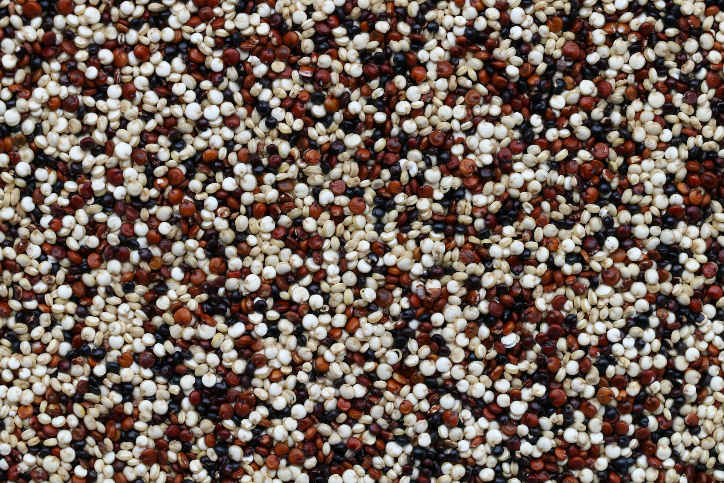
1 cup Quinoa = 8g
1/2 cup Beans (Kidney, Black) = 8g
1/2 cup of Hummus = 9.5g
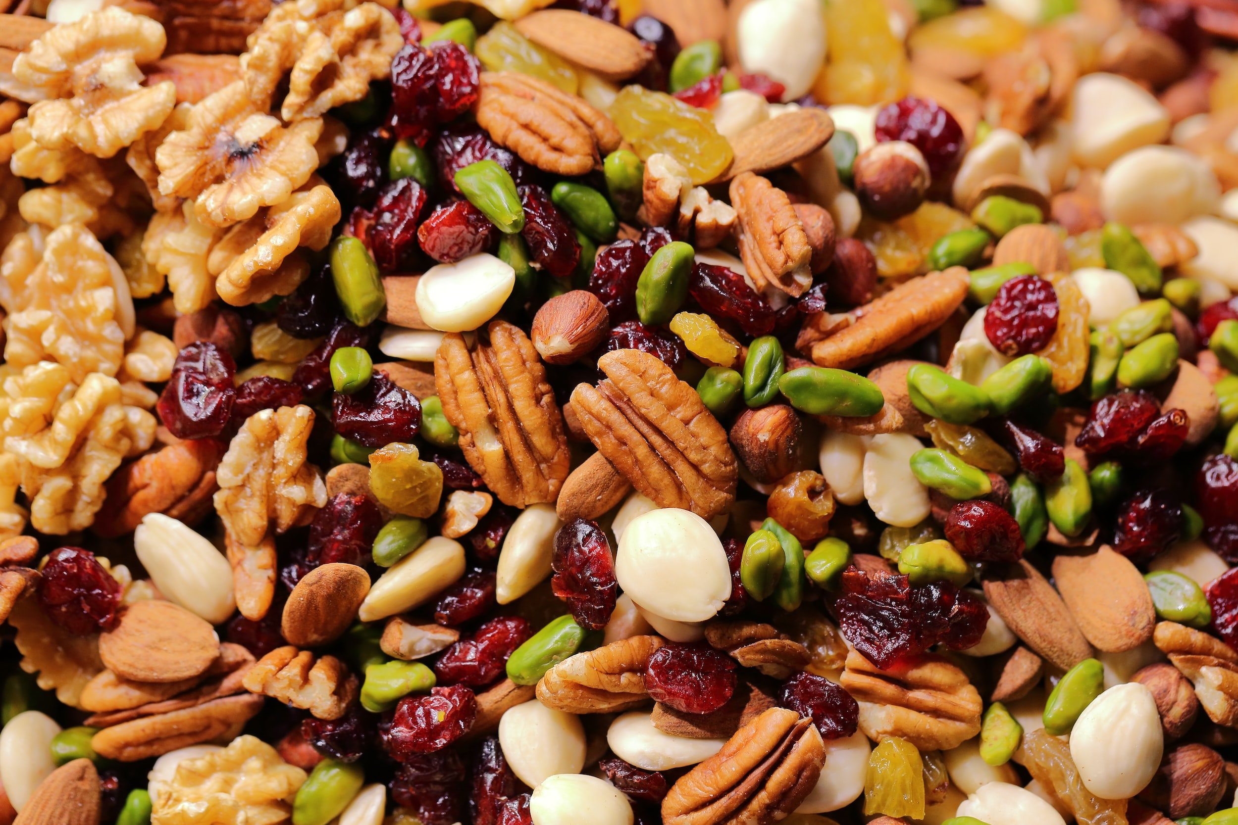
1/2 cup of Nuts = 13.5g
3 oz Beef, Fish or Pork = 21g
3 oz Chicken Breast = 26g


3 Daily Meals Balanced Protein Recommendations (1-4)
-100lbs = 23g/meal
-120lbs = 27g/meal
-140lbs = 32g/meal
-160lbs = 36g/meal
-180lbs = 41g/meal
-200lbs = 45g/meal
-220lbs = 50g/meal
5 Daily Meals Balanced Protein Recommendations (1-4)
-100lbs = 14g/meal
-120lbs = 16g/meal
-140lbs = 19g/meal
-160lbs = 22g/meal
-180lbs = 24g/meal
-200lbs = 27g/meal
-220lbs = 30g/meal
Protein intake Post Exercise (2)
15-25g
0-2 hours post exercise
Food based proteins preferred
When not available, 3rd party tested protein powder supplements can provide a practical alternative
How to Find out Protein levels?
Protein Helps to Counteract Anabolic Resistance
Older athletes can develop Anabolic Resistance, which is a slowed response to protein intake and strength training. (4-6)
We all lose muscle mass as we age, which is termed sarcopenia. Sarcopenia typically affects our type II or fast twitch muscles fibres, beginning in our 40s-50s and advances annual thereafter. (4,6,7)
Sarcopenia can be accelerated during periods of muscle disuse or unloading from injures, illnesses or hospitalizations.
As we age the consequences of sarcopenia include an increased risk of developing chronic diseases, falls, fractures, loss of independence and death.
It appears that sarcopenia is actually due to anabolic resistance. Interestingly anabolic resistance may be mostly attributed to reduced physical activity levels and lower protein intake in the older population (5,6)
Since aging is associated with anabolic resistance, older athletes require greater relative protein intakes to maintain muscles and don’t respond as quickly to strength training. (3-5)
Unfortunately what typically happens is: (4)
- 8g Breakfast (Goal = 20-30g)
-12g Lunch (Goal = 20-30g)
-40g Dinner (Goal = 20-30g)
That’s why it’s important for older athletes to ingest a balanced amount of protein (20-30g) across a minimum of 3 meals a day, along with strength training 2-3 times / week. (3-5)
-
1) Desbrow, B. (2021). Youth Athlete Development and Nutrition. Sports Med, 51 (Suppl 1), 3-12
2) Thomas D. T., Erdman, K. A., & Burke, L. M. (2016). American College of Sports Medicine Joint Position Statement. Nutrition and Athletic Performance. Med Sci Sports Exerc, 48(3), 543-568
3) Morton, R. W., Murphy, K. T., McKellar, S. R., Schoenfeld, B. J., Henselmans, M., Helms, E., et al. (2018). A systematic review, meta-analysis and meta-regression of the effect of protein supplementation on resistance training-induced gains in muscle mass and strength in healthy adults. Br J Sports Med, 52(6), 376-384
4) Breen, L., & Phillips, S. M. (2011). Skeletal muscle protein metabolism in the elderly: Interventions to counteract the 'anabolic resistance' of ageing. Nutrition & metabolism, 8(68), 1-11
5) Moore, D. R., Churchward-Venne, T. A., Witard, O., Breen, L., Burd, N. A., Tipton, K. D., & Phillips, S. M. (2015). Protein ingestion to stimulate myofibrillar protein synthesis requires greater relative protein intakes in healthy older versus younger men. Gerontology, 70(1), 57–62
6) Wall, B. T., Gorissen, S. H., Pennings, B., Koopman, R., Groen, B. B., Verdijk, L. B., & van Loon, L. J. (2015). Aging Is Accompanied by a Blunted Muscle Protein Synthetic Response to Protein Ingestion. PloS one, 10(11), e0140903
7) Grosicki, G. J., Zepeda, C. S., & Sundberg, C. W. (2022). Single muscle fibre contractile function with ageing. Physiology, 600(23), 5005–5026
How to Stretch
Mobility is defined as the ability to voluntarily move a joint through its full range of motion (ROM). Your mobility depends on individual anatomical and physiological components such as: (1, 2)
Muscles and tendons
The states of ligaments, bones and cartilages that form the joint
Spinal reflex activity
Stretch tolerance
Stretching your muscles is an effective way to increase mobility. (1, 2)

In order to improve mobility with stretching: (1-4)
Hold each stretch for 2-5min
Intensity of 30-40% of maximal tolerable stretch
Frequency of 5 days minimum per week
It’s important to note that stretching has just been shown to improve mobility. Stretching will NOT help to: (5-7)
Enhance sports performance
Improve health
Prevent sports injuries
Reduce muscle soreness after physical activity
-
1) Behm, D. G., Alizadeh, S., Anvar, S. H., Drury, B., Granacher, U., & Moran, J. (2021). Non-local Acute Passive Stretching Effects on Range of Motion in Healthy Adults: A Systematic Review with Meta-analysis. Sports medicine, 51(5), 945–959.
2) Thomas, E., Bianco, A., Paoli, A., & Palma, A. (2018). The Relation Between Stretching Typology and Stretching Duration: The Effects on Range of Motion. International journal of sports medicine, 39(4), 243–254.
3) Nakamura, M., Ikezoe, T., Takeno, Y., & Ichihashi, N. (2013). Time course of changes in passive properties of the gastrocnemius muscle-tendon unit during 5 min of static stretching. Manual therapy, 18(3), 211–215.
4) Wyon, M., Felton, L., & Galloway, S. (2009). A comparison of two stretching modalities on lower-limb range of motion measurements in recreational dancers. Journal of strength and conditioning research, 23(7), 2144–2148.
5) Lauersen, J. B., Bertelsen, D. M., & Andersen, L. B. (2014). The effectiveness of exercise interventions to prevent sports injuries: a systematic review and meta-analysis of randomised controlled trials. British journal of sports medicine, 48(11), 871–877.
6) Nuzzo J. L. (2020). The Case for Retiring Flexibility as a Major Component of Physical Fitness. Sports medicine, 50(5), 853–870.
7) Herbert, R. D., de Noronha, M., & Kamper, S. J. (2011). Stretching to prevent or reduce muscle soreness after exercise. The Cochrane database of systematic reviews, (7), CD004577.